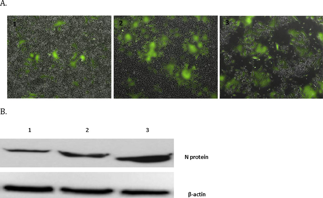
Figure 3.

MV-GFP infection kinetics in the moderately susceptible 143B sarcoma cell line. MV-GFP infection leads to (A) GFP expression is increased over time until monolayer obliteration. Images A1, 2, 3 were taken on days 1, 2, and 3 respectively following infection (4 × magnification). (B) Increased expression of measles N protein during the same time course.
