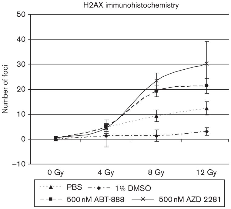
Fig. 2.
Number of foci detected using laser confocal microscopy and fluorescent Fluor 647 anti-H2A.X-phosphorylated (Ser139) antibody. Double-stranded breaks (red) are clearly augmented in cells incubated with 500 nmol/l of ABT-888 and 500 nmol/l of AZD-2281 compared with PBS and 1% dimethyl sulfoxide controls. Image analysis was performed using ImageJ and the ‘analyze particle’ function.

