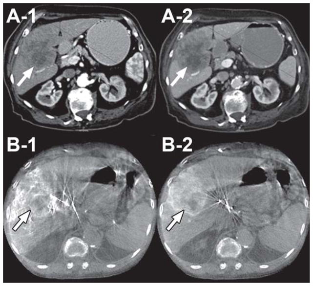
Figure 1.
70-year-old female with poorly differentiated carcinoma (Child-Pugh score = 7/Class B, no ascites, no cirrhosis) in liver segments 5 and 6 (arrows). A-1 and A-2 are arterial and venous phase contrast-enhanced CT images, respectively. B-1 and B-2 are early and late arterial dual-phase CBCT images, respectively, immediately before lipiodol treatment. Note how the late arterial phase image (B-2) shows very clear tumor parenchymal space, border, and matching with the CT image (A-2).

