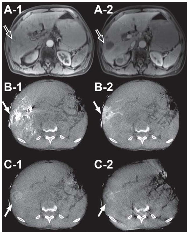
Figure 2.
59-year-old male patient with HCC (Child-Pugh score = 6/Class A, no ascites, cirrhosis present) in liver segments 2, 4a, 7, and 8 (arrow outlines). A-1 and A-2 are pre-intervention contrast enhanced MR images at 20 and 70 sec post-contrast injection, respectively. B-1, -2 and C-1, -2 are pre- and post-drug eluting bead administration, respectively, of early (B-1 & C-1) and late (B-2 & C-2) arterial dual-phase CBCT images. Note the change in tumor enhancement and lack of feeding vessel enhancement before and after bead embolization for both early and late arterial images (solid arrows) in the axial slices, indicating successful embolization.

