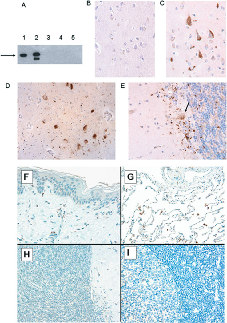
Figure 3. Tissue distribution of PDE4A8 protein.
(A) Immunoblotting of lysates from COS-7 cells transfected with a plasmid expressing PDE4A8. The recombinant protein contained a VSV epitope at its C-terminus. Lanes were immunoblotted as follows: 1, anti-VSV antibody; 2, anti-PDE4A8 antibody; 3, pre-immune serum; 4, anti-PDE4A8 antibody, cells transfected with vector only; and 5, anti-PDE4A8 antibody plus peptide used as immunogen (62.5 µg/ml). The recombinant protein migrated at 125 kDa (arrow). The less-intense band seen in lane 2 was thought to be a proteolytic fragment; we have observed such fragments before when PDE4 proteins are expressed at high levels in COS-7 cells [25]. (B) Immunohistochemistry of human cerebral cortex, using pre-immune serum. (C)–(I) show immunohistochemistry of human tissues, using the anti-PDE4A8 antibody. The tissues are as follows. (C) Cerebral cortex, showing several pyramidal neurons that stained strongly. (D) Spinal cord, showing a cluster of anterior horn neurons that stained strongly. (E) Cerebellar cortex; staining was seen in a diffuse band at the base of the Purkinje cell layer, including Purkinje cells (arrow). Staining of Purkinje cell bodies was most pronounced in a perinuclear pattern, extending into dendrites that project into the deeper portions of the molecular layer. (F) Skin, showing no staining. (G) Lung, showing staining of some rare scattered pulmonary macrophages, but no staining of alveolar or interstitial cells. (H) Ovarian parenchyma, showing no staining. (I) Germinal centre of a benign lymph node, showing no staining. All histological sections were at an original magnification of 400×.

