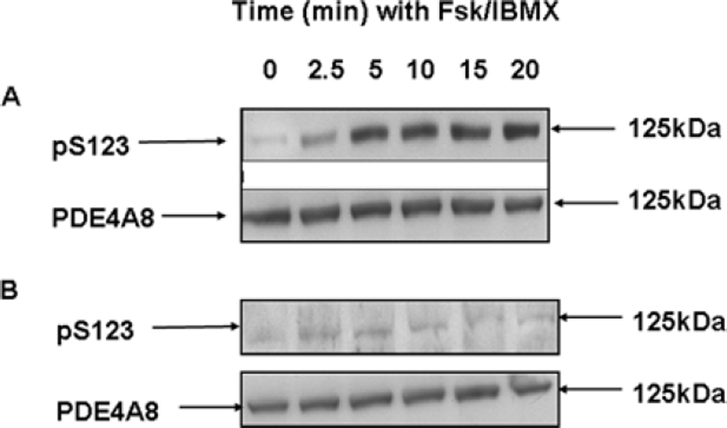
Figure 5. Phosphorylation of PDE4A8 by PKA.
(A) COS-7 cells transfected to express PDE4A8 were treated with IBMX and forskolin (Fsk) to increase intracellular cAMP levels and activate PKA. At the indicated times after treatment, the cells were lysed and the lysates subjected to SDS/PAGE and immunoblotted. In the upper panel, they were immunoblotted with an antibody specific for the serine residue phosphorylated by PKA [i.e. Ser123 (pS123) in PDE4A8; Figures 1A and 1B]. In the lower panel, they were immunoblotted with the pan-4A antibody, to ensure that equal amounts of PDE4A8 protein were loaded on to each lane. (B) As in (A), but also with treatment with the PKA inhibitor H89. PDE4A8 migrated as a 125 kDa band. These results were typical of those obtained at least three times.

