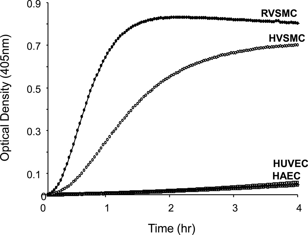
Figure 2. Comparison of PK activation by endothelial and VSMC.
Rat aortic VSMCs, human aortic VSMCs (HVSMCs), human umbilical vein endothelial cells (HUVECs) and human aortic endothelial cells (HAECs) were subcultured in different wells and incubated with 3.58 nM of PK and 0.4 mM of S-2302. Optical densities were recorded at 405 nm at every 2 min for 2 hrs. While HUVECs and HAECs did not show PK activation, rat VSMCs (closed circle) and HVSMCs (open circle) showed remarkable activation of PK in the absence of FXII. Graphs are from a representative set of 3 experiments.

