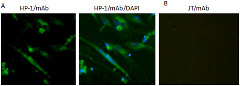
Fig 1. DHAV-1 HP-1- and DHAV-3 JT-infected DEF cells detected with mAb 2D10 by IFA.

(A) DHAV-1 HP-1-infected DEF cells; Infected cells were stained with mAb 2D10 and then with an FITC-conjugated goat anti-mouse antibody, and finally with DAPI. Stained cells were visualized by means of fluorescence microscopy. (magnification 300×). (B) JT-infected DEF or uninfected DEF cells detected by mAb 2D10 (negative control).
