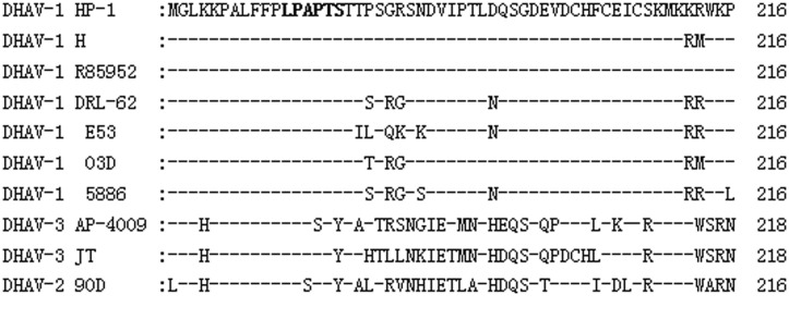
Fig 6. Sequence alignment of 10 DHAV strains around the epitope-coding region of the VP1 protein.

Amino acid positions for each individual sequence are numbered on the right. DHAV-1 HP-1 strain sequences are shown at the top; dashes indicate identical amino acids. The identified epitope is in bold.
