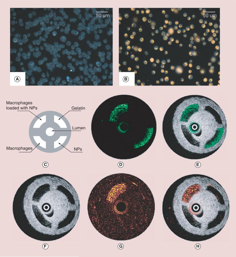
Figure 7. Darkfield reflectance optical images of intact murine macrophages.
(A) Murine macrophages loaded with gold nanoparticles (B). The diagram (C) and the IVUS image (F) of the tissue mimicking phantom. The dynamic ranges of IVUS and IVPA images were 50 and 17 decibel, respectively. The IVPA images of the same cross-section of the phantom were taken at 532 nm (D) and 680 nm wavelength (G). The combined IVUS and IVPA images of the phantom (E; 532 nm wavelength and H; 680 nm wavelength) indicate the origin of the PA responses in IVPA images.
IVPA: Intravascular photoacoustic; IVUS: Intravascular ultrasound; NP: Nanoparticle.

