
Figure 1.
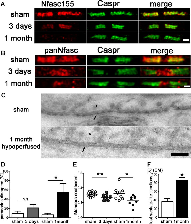
Paranodal disruption occurs early in response to hypoperfusion in the corpus callosum. A, B, Colocalization of axonal Caspr and glial Neurofascin protein indicates intact septate-like junctions at the paranodes in the sham group. In response to hypoperfusion, at both 3 d and 1 month, there is a selective loss of Neurofascin colocalization with Caspr, which is indicative of a disruption of the paranodes. Scale bar, 1 μm. C, Electron micrographs show paranodal disruption in response to hypoperfusion in the optic nerve. Arrows indicate axonal membrane at the paranodal region; asterisks indicate paranodal loop. Disruption of septate-like junctions is indicated by loss of transverse bands. Scale bars, 0.1 μm. D, There is a nonsignificant increase in the number of paranodes disrupted at 3 d hypoperfusion (n = 5) compared with shams (n = 5), which is significant at 1 month after hypoperfusion (n = 4) compared with shams (n = 6). Analysis was conducted in the corpus callosum. *p < 0.05 (unpaired t test, two-tailed). E, The overlap coefficient after Manders, which is insensitive to differences in signal intensities between the two channels, shows significant loss in colocalization after 3 d and after 1 month of hypoperfusion (3 d sham: n = 13, hypoperfusion: n = 12; 1 month sham: n = 9, hypoperfusion: n = 9). Analysis was conducted in the corpus callosum. *p < 0.05, **p < 0.005 (unpaired t test, two-tailed). F, A significant increase in paranodes without septate-like junctions was observed after 1 month of hypoperfusion. *p < 0.05 (unpaired t test, two-tailed).