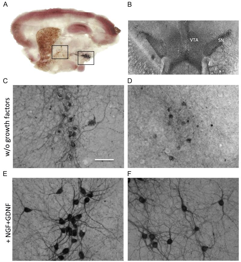
Fig. 1.
A sagittal vibrosection (A) shows cholinergic and dopaminergic neurons stained by immunohistochemistry for choline acetyltransferase (brown) or tyrosine hydroxylase (black), respectively. This vibrosection was counterstained with calbindin (red) to show the brain structures. A coronal vibrosection (B) shows tyrosine-hydroxylase positive dopaminergic neurons of the SN and VTA. When vibrosections were incubated without growth factors, only a few not healthy cholinergic nBM (C) or dopaminergic SNc (D) neurons could be observed. However, when vibrosections were incubated with 10 ng/ml NGF and GDNF the number of healthy cholinergic (E) and dopaminergic (F) neurons markedly increased. Scale bar in C=1800 μm (A), 930 μm (B), 80 μm (C–F).

