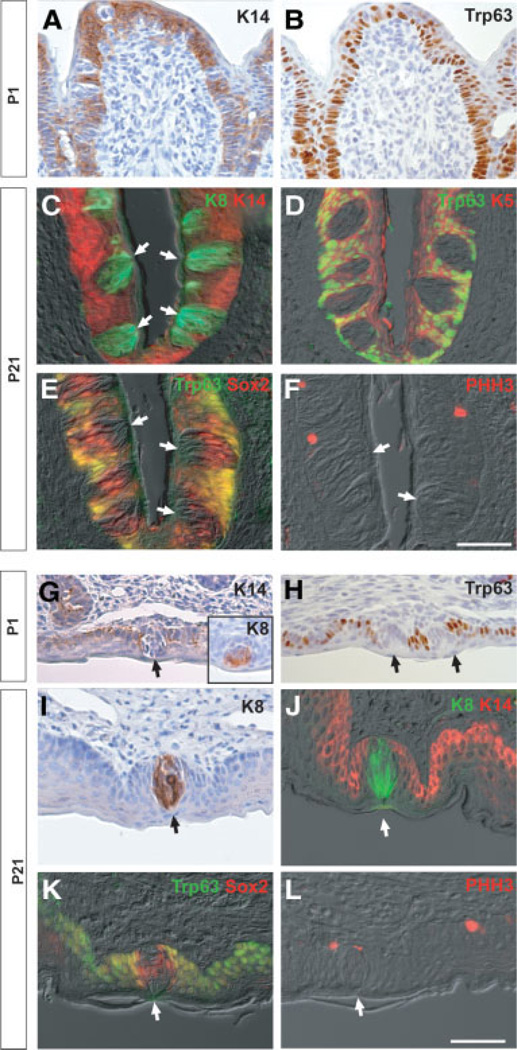
Figure 2.
Expression of epithelial markers in the circumvallate papilla and soft palate at different stages. (A–F): Sections of circumvallate papilla at P1 (A, B) and P21 (C–F). Shown are the expression of keratin 14 (K14) (A) and Trp63 (B) at P1 and the expression of K8/K14 (C), Trp63/K5 (D), Trp63/Sox2 (E), and phosphohistone H3 (PHH3) (F) merged with differential interference contrast (DIC) images. Note the exclusion of K14, Trp63, and K5 from the interior of the taste bud (TB), which contains K8+ cells with a sharp boundary in perigemmal cells (C, D). Note also the coexpression of Sox2 and Trp63 in cells around the TBs (E). (G–L): Sections of soft palate at P1 (G, H) and P21 (I–L). Shown are expression of K14 (G), K8 (G, inset), and Trp63 (H) at P1 and expression of K8 (I), K8/K14 (J), Trp63/Sox2 (K), and PHH3 (L) merged with DIC images. Note again the sharp boundary between K8+ and K14+ cells and the coexpression of Trp63 and Sox2 in perigemmal cells adjacent to the TBs (J, K). Arrows indicate TBs. Scale bar, 50 µm. Abbreviation: P, postnatal day.

