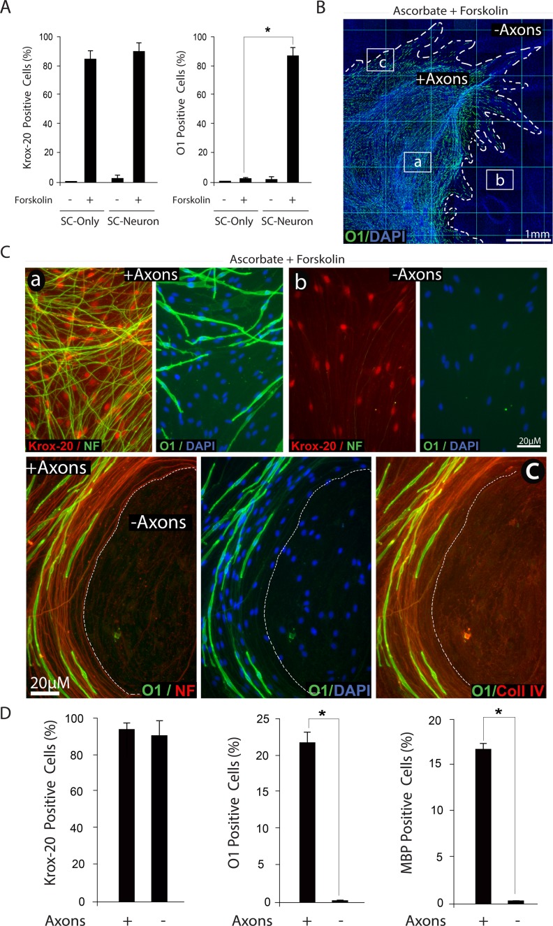
Fig 10. Induction of Krox-20, O1 and MBP expression by combined administration of forskolin and ascorbate: dependency on axonal contact.
Experiments in SC-neuron cultures were identical to those described in Fig. 8, with the exception of experiments shown in panel A, where the responses of isolated (SC-only) and axon-related SCs (SC-neuron) were compared at 3 days post-stimulation. Representative areas located within (a), outside (b) and at the frontier (c) of the axonal outgrowth are shown at high magnification to reveal details of the morphology of the cells, their relationship to axons (neurofilament staining, NF) and their expression of Krox-20, O1 and collagen type IV, as indicated. In panel B, a low magnification image of a representative SC-neuron culture treated with ascorbate and forskolin is shown to provide a reference to the relative location of the areas displayed in C. Note the selective distribution of O1 positive cells (green) with respect to the total number of cells (DAPI) and the extension of the neurite outgrowth (dotted lines). A quantification of the percentage of Krox-20, O1 and MBP positive cells in selected areas within (+ axons) and outside (- axons) the axonal outgrowth is provided in panel D. Whereas Krox-20 expression was enhanced throughout the culture system, the expression of O1 and MBP was confined to those SCs that physically interacted with axons (C). Note that even those SCs that do not contact axons exhibit a profile of extracellular collagen IV expression (C, lower panels).

