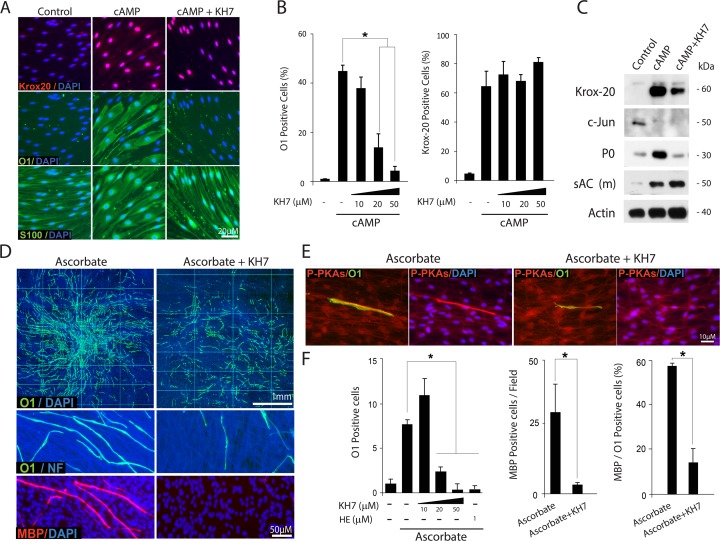
Fig 12. Inhibition of O1 expression and morphological differentiation by pharmacological sAC antagonists.
The experimental design and analysis of results carried out using SC-only (A-C) and SC-neuron cultures (D-F) were identical to those of previous figures. In panels A-C, isolated SCs were left untreated (control) or treated with cAMP (CPT-cAMP, 250 μM) in the absence or presence of increasing doses of KH7, as indicated. In panels D-F, SC-neuron cultures were stimulated with ascorbate-containing medium in the absence (control) or presence of KH7, which was used at 20 μM unless otherwise stated in the figure. Cultures were double-immunostained with antibodies against O1 and MBP (D) or O1 and P-PKA substrates (E). Data presentation and quantitative analysis of Krox-20, O1 and MBP expression was done as previously described. In panel F, a condition that used 2-hydroxy-estradiol (HE) was included in the quantitative analysis of O1 expression for confirmation of results. KH7 abrogated the morphological transformation associated with the differentiation of isolated (A, S100 expression) and axon-related SCs (D-E, O1 expression). KH7 diminished the total levels of expression of O1 (A-B and D-F), P0 (C) and MBP (D-F) without reducing the expression of Krox-20 (A-C) or increasing that of c-Jun (C).

