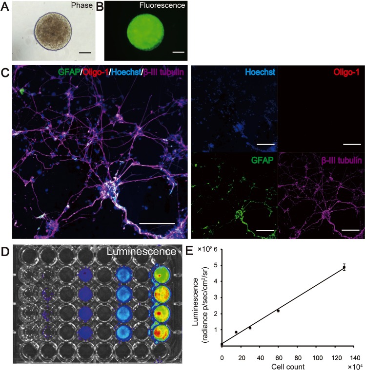
Fig 1. Detection of bioluminescence and fluorescence signals in lentivirally transfected 253G1-NS/PCs in vitro.
Phase-contrast (A) and fluorescence (B) images of a neurosphere derived from tumorigenic 253G1 induced pluripotent stem cells. Neural stem/progenitor cells (NS/PCs) differentiated into β-III tubulin-positive neurons and glial fibrillary acidic protein (GFAP)-positive astrocytes in vitro (C). Bioluminescence imaging was used to detect bioluminescence signals in various numbers of 253G1-NS/PCs (0, 1.5 × 10⁵, 3 × 10⁵, 6 × 10⁵, and 1.2 × 10⁶ cells per well) (D). A direct linear correlation was found between cell numbers and photon counts in vitro (E). Scale bars in A–C, 1,000 μm.

