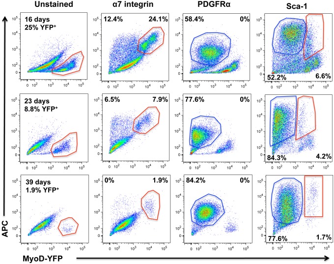
Fig 4. Flow cytometry profiles of myosphere-derived cells isolated from YFP-MyoD mice.
Shown are flow cytometry profiles of myosphere-derived cells isolated from YFP-MyoD mice that had remained in culture for 16 (upper panels), 23 (middle panels), and 39 days (lower panels). Shown are the flow cytometry profiles for unstained cells and for cells incubated with APC antibodies against α7-integrin, PDGFRα, and Sca-1. Y-axis shows APC expression and the x-axis YFP expression (MyoD).

