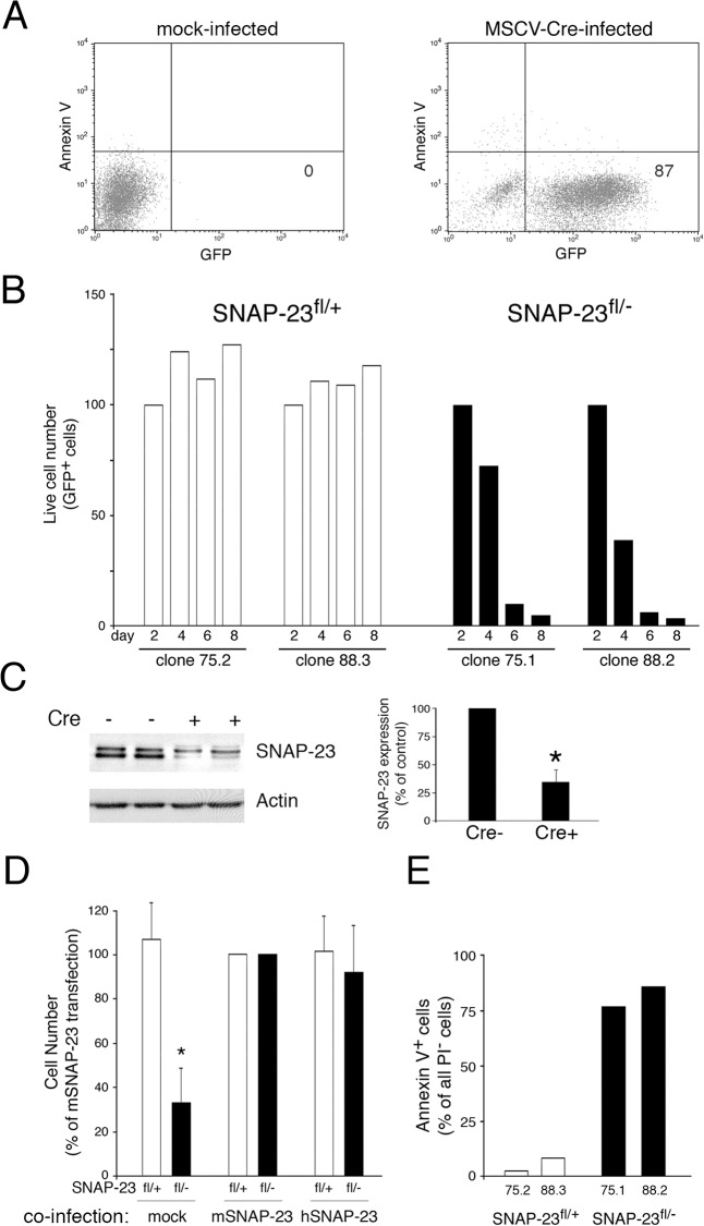
Fig 6. Deletion of SNAP-23 leads to acute death of MEFs.
MEF lines were generated from SNAP-23fl/+ mice (clones 75.1 and 88.2) or SNAP-23fl/- mice (clones 75.2 and 88.3). (A) Infection of a typical SNAP-23fl/+ MEF culture infected with empty retrovirus (left panel) or GFP-Cre retrovirus (right panel) shows that two days after infection the vast majority of MEFs were viable (Annexin V-) and expressed GFP-Cre. (B) The indicated MEF lines were infected with GFP-Cre-expressing retrovirus and the number of live cells present in each culture (based on staining with PI) was determined at different times. The absolute cell recovery in each condition was expressed relative to the amount of cells present two days after infection (control experiments showed that there was no Cre-dependent cell death in any line after only two days of infection). The data shown are representative of two independent experiments analyzed at day 2, 4, 6, 8 and one experiment analyzed at day 1, 3, 5, 7. (C) Adherent SNAP-23fl/- MEFs were isolated 4 days after retroviral transduction with Cre (Cre+) or after mock-transduction (Cre-). Equal numbers of cells from each culture were analyzed by SDS-PAGE and immunoblotting using a SNAP-23 antibody. The blot was re-probed for anti-β actin mAb as a loading control. The amount of SNAP-23 present in each cell lysate was normalized to the amount of actin present and the data shown are mean +/- SD of three independent experiments (*p<0.05). (D) MEF lines generated from SNAP-23fl/+ mice or SNAP-23fl/- mice were co-infected with retrovirus containing GFP-Cre and retrovirus containing nothing (mock), mouse SNAP-23 (mSNAP-23), or human SNAP-23 (hSNAP-23). After 4 days the number of live cells present in each culture (based on staining with PI) was determined and was expressed relative to the number of cells recovered in the mSNAP-23 co-infection condition. The data shown are mean +/- SD of four independent experiments (*p<0.05). (E) The indicated MEF lines were infected with GFP-Cre-expressing retrovirus and after four days the cells were stained with PI and Annexin V and analyzed for GFP-Cre expression. The percentage of viable (PI-) GFP-Cre+ Annexin V+ cells in each culture was determined by flow cytometry. The data shown are representative of three independent experiments.

