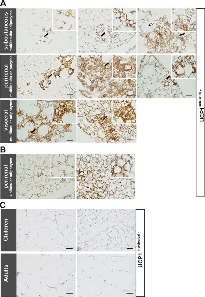
Fig 1. Immunohistochemical evidence of brown-like adipocytes in adipose tissue samples.
A: Immunohistochemical staining of AT samples showed multilocular adipocytes, which stained positive for UCP1 (black arrows), surrounded by UCP1-negative white unilocular fat cells (white arrows) in subcutaneous (top), perirenal (middle) and visceral (bottom) AT samples of children. B: Two perirenal unilocular samples also exhibited positive UCP1 staining (grey arrows). C: Representative images of UCP1-negative subcutaneous (top, left) and perirenal (top, right) AT samples of lean children and subcutaneous samples of lean adults (bottom). Nuclei were counterstained with Mayer’s hematoxylin. Scale bars represent 50 μm in each panel.

