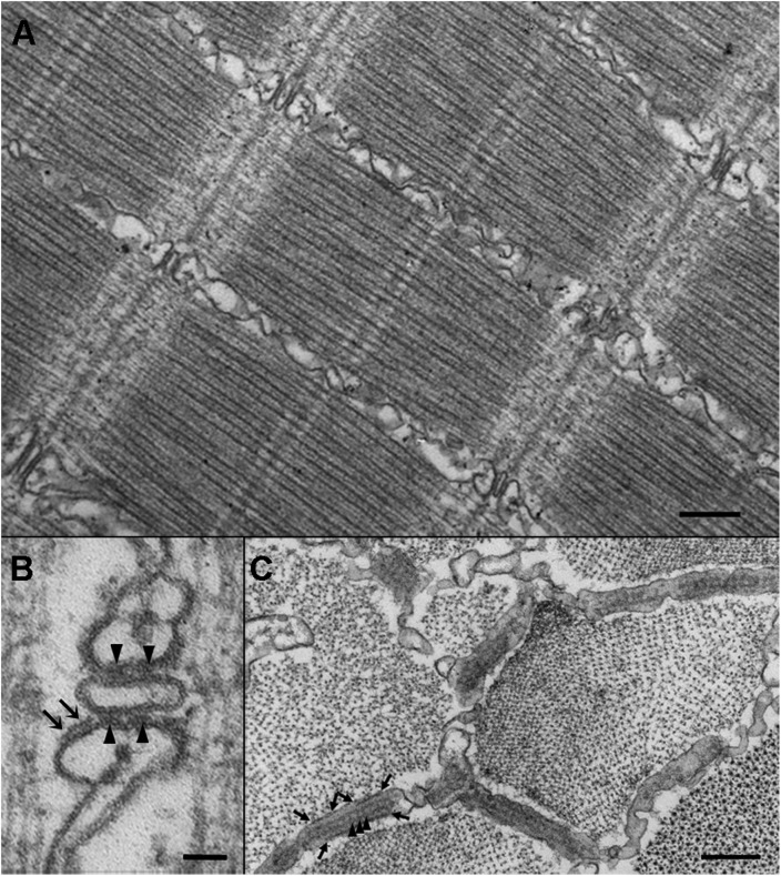
Figure 1.
Images from thin sections of tail myotomes from 72 hpf larvae. (A) Detail of a muscle fiber in a 72 hpf larva. At this age, fibers in proximal myotomes have well-differentiated myofibrils and CRUs in the form of triads located at the Z-line. Bar, 250 nm. (B) Triads in fast-twitch fibers, constituting the majority of the tail, have two sets of “feet”: JF located between SR and T-tubule membranes (arrowheads), and PJF located near the junction but on the side of the SR facing the myofibrils (arrows). Bar, 50 nm. (C) Cross sections of the fibers offer grazing views of the junctional gap, occupied by two rows of JF (arrowheads) and two other discontinuous rows of PJF (delimited by the arrows) on the side of the SR cisterna. Bar, 250 nm.

