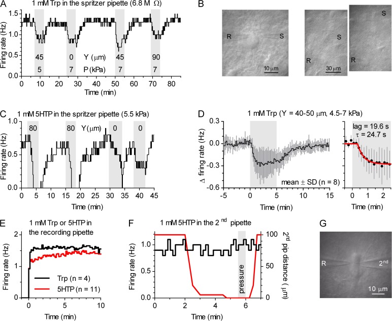
Figure 12.
5-HT–mediating suppression of firing originates in surrounding neurites. (A) Time course of representative experiment in which 1 mM Trp was spritzed 45 µm lateral, 90 µm lateral and directly toward the recorded neuron. (B) Photographs showing recorded neuron, recording pipette (R), and spritz pipette (S) arrangement for the experiment shown in A. (C) Time course of a representative experiment in which 1 mM 5HTP was spritzed 80 µm lateral and directly toward the cell body of the recorded neuron. Transient decrease in the firing rate at the end of the cell body–directed pulses is an artifact of tissue movement. (D) Average time course for experiments in which 1 mM Trp was spritzed 40–50 µm lateral to the recorded neuron (n = 8). Symbols represent the mean ± SD of the normalized, binned firing rate, expressed as absolute difference in the baseline firing rate computed over the 5 min preceding Trp application in each experiment. Shaded area corresponds to the spritz duration. Right, the onset of Trp-induced change on an enlarged scale. Red curve is a data fit of means with the function of the form Y = {0, if t < lag; YTrp(−1 + e−(t − lag)/τ), if t ≥ lag}, where lag is time from the application of Trp to the beginning of change, τ is the exponential time constant, and YTrp is the change in firing rate when steady-state level is achieved (lag = 19.6 s; 13.8–25.3 s 95% C.I.; τ = 24.7 s; 17.7–40.8 s 95% C.I.; YTrp = 0.27 Hz; 0.26–0.29 Hz 95% C.I. Hz; R2 = 0.969). (E–G) Direct somatic application of Trp and 5HTP with the recording pipette did not change the firing rate of the recorded serotonergic neuron. (E) Average time course for loose-seal cell-attached recordings in which 1 mM Trp or 1 mM 5HTP was included in the recording pipette solution. (F) Time course of an experiment in which the recorded neuron was rapidly approached with the second, nonrecording wide-tip patch pipette (Rpip = 1.9 MΩ) containing 1 mM 5HTP during recording with 5-HT precursor-free pipette solution. The red line represents the distance between tip of the pipette and the neuron. Positive pressure was applied to the 5HTP-containing pipette for 30 s, indicated by the shaded area. (G) Photograph of experiment in F taken during the period when both the recording (R) and the second (second) pipette were touching the recorded neuron.

