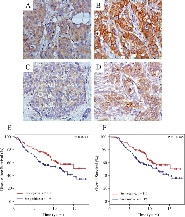
Fig 6. Immunohistochemical staining of Src expression and phosphorylation in primary breast tumors and Kaplan-Meier survival estimates relative to the expression of Src at the plasma membrane.
Representative pictures of weak (A) and strong (B) immunohistochemical staining of total Src and of weak (C) and strong (D) immunohistochemical staining of phosphorylated Src (Tyr416) expressed in primary breast tumors. Kaplan-Meier survival plots demonstrating percentage disease-free (E) and overall (F) survival in patients with tumors with or without Src expressed at the plasma membrane.

