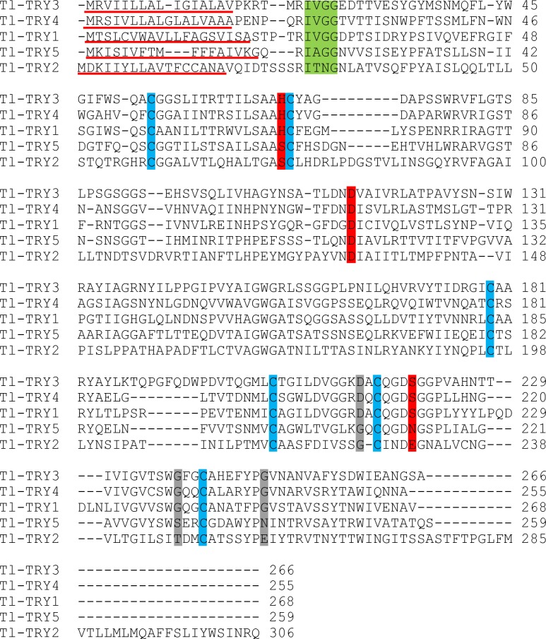
Fig 7. ClustalW2 alignment of T. licus licus trypsin-like proteins.
Many sequences preserved the characteristics of digestive serine proteases. The signal peptide is shown with a red underline. The cleavage site is shown in a green box. Red boxes indicate the active site residues. Grey boxes indicate the substrate binding region and cysteines that are possibly involved with disulfide bonds are shown in blue.

