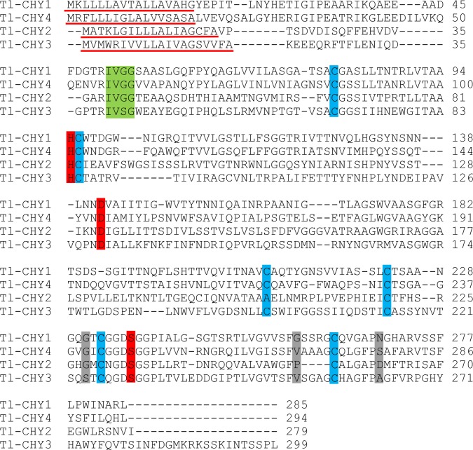
Fig 8. ClustalW2 alignment of T. licus licus chymotrypsin-like proteins.
Many sequences preserved the characteristics of digestive serine proteases. The signal peptide is shown with a red underline. The cleavage site is shown in green box. Red boxes indicate the active site residues. Grey boxes indicate the substrate binding region and cysteines that are possibly involved with disulfide bonds are shown in blue.

