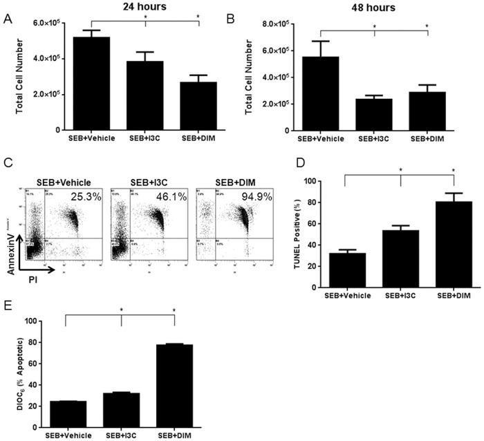
Fig 3. Treatment with I3C or DIM induces apoptosis in SEB-activated cells.
Splenocytes from C57BL/6 mice were activated with SEB (1μg/ml) in vitro in 96-well plates in complete culture media in the presence or absence of I3C or DIM (100μM). Cells were collected and counted to determine cell number (A-B). Representative plots of 24 hour cultures stained with Annexin V/PI are shown (C). In addition, cells were stained for TUNEL after 24 hour culture and analyzed by flow cytometry to detect apoptosis (D). The cells were also stained with DiOC6 to determine if the observed apoptosis was mediated through the mitochondrial pathway (E). Statistical significance (p-value <0.05) was determined using GraphPad Prism analysis software with one-way ANOVA and Tukey’s multiple comparison test (* indicates significance compared to SEB+Vehicle).

