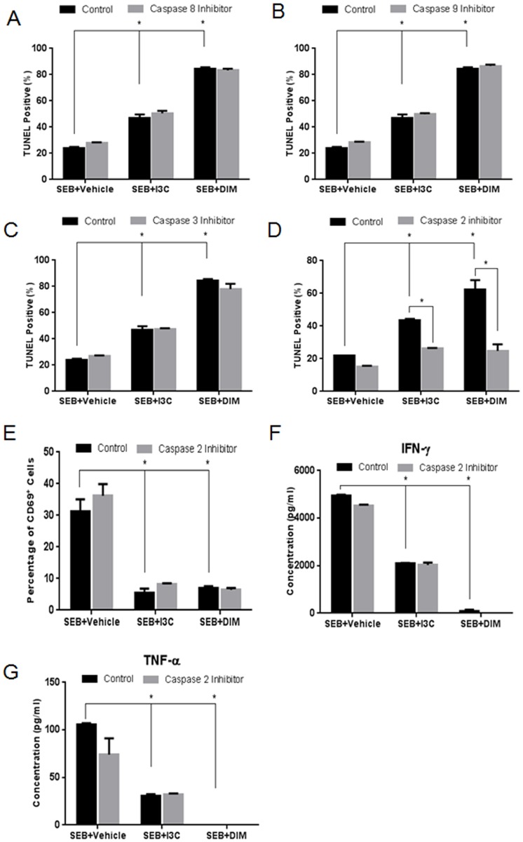
Fig 4. Role of caspase-2 in I3C and DIM-mediated apoptosis, cell activation, and cytokine release.
Splenocytes from C57BL/6 mice were cultured and activated with 1μg/ml of SEB, in the absence or presence of I3C or DIM (100µM). The cultures were treated with 100µM of inhibitors for caspases-8 (A), -9 (B), -3 (C), or-2 (D). Percentage of TUNEL positive cells was determined using a TUNEL kit and analyzed by flow cytometry. Percentage of CD69 expressed in experimental groups was determined in the presence or absence of caspase-2 inhibitor (E). ELISAs were used to detect cytokine concentrations from supernatants collected from 24-hour cultured experimental groups treated with or without capase-2 inhibitor for IFN-γ (F) and TNF-α (G). Statistical significance was determined using GraphPad Prism analysis software with one-way ANOVA and Tukey’s multiple comparison to test between different groups. Two-way ANOVA and Sidak’s multiple comparison test were used to compare controls with those containing inhibitors. Significance is indicated by * and has a p-value <0.05.

