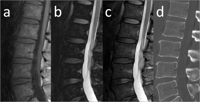
Fig 3. MRI of the lumbar spine in a false-positive case misclassified on the basis of β2-microglobulin.

Sagittal MR images and sagittal reconstructed CT of a 53-year-old man with asymptomatic myeloma and a fat-signal fraction of 64% are shown. Serum creatinine level was 2.4 mg/dl, β2-microglobulin was 5.99 mg/l, and BMPC% from the iliac crest was 7.5%. The elevated β2-microglobulin level might have resulted from renal impairment. a) T1-weighted imaging shows diffusely decreased signal intensity in bone marrow. Bone marrow signal intensity is almost equal to that of intervertebral disc. b) Calculated out-phase image. c) Calculated in-phase image. d) Sagittal reconstructed CT.
