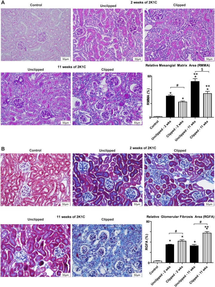
Fig 2. Histological analysis of kidney sections 2 and 11 weeks after renal artery clip placement.
A) PAS staining of control, 2K1C unclipped and 2K1C clipped kidneys 2 and 11 weeks after clip placement. B) Masson’s Trichrome staining of control, 2K1C unclipped, and 2K1C clipped kidneys 2 and 11 weeks after clip placement. *p < 0.001 vs. Control, p** < 0.001 vs. 2 wks, #p < 0.001, $p < 0.01.

