Figure 7.
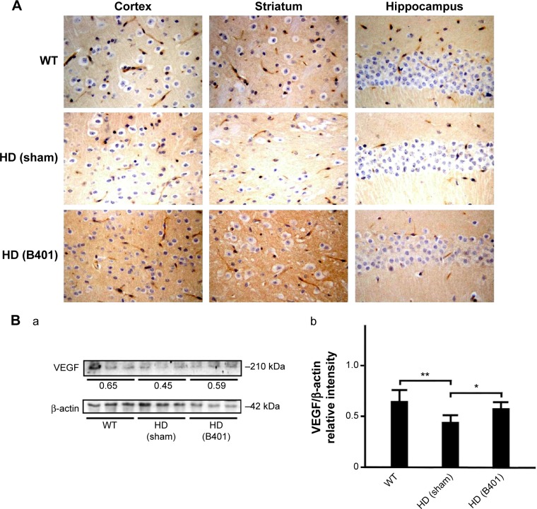
VEGF levels in the brain tissue of 2-month R6/2 mice were upregulated under oral B401 treatment.
Notes: (A) Immunohistochemical staining illustrates expression levels of VEGF in the cortex, striatum, and hippocampus of 2-month R6/2 mice and their WT. Expression levels of VEGF in the brain tissue of 2-month R6/2 mice were visibly enhanced under oral B401 treatment. The width × height of each figure is 150 μm ×100 μm. (B) Western blotting analysis shows the following: (a) expression levels of VEGF in whole brain tissue of 2-month R6/2 mice and their WT and (b) quantified brain VEGF levels of 2-month R6/2 mice (HD sham, n=6) were significantly reduced than their WT (n=6), and were significantly increased under oral B401 treatment (HD B401, n=6). Values are mean ± SEM (*P<0.05, **P<0.01, one-way ANOVA followed by a Student–Newman–Keuls multiple comparisons posttest).
Abbreviations: WT, wild type; HD, Huntington’s disease; VEGF, vascular endothelial growth factor; SEM, standard error of the mean; ANOVA, analysis of variance.

