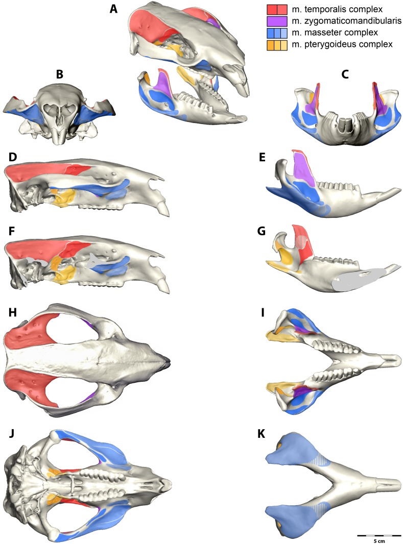
Fig 1. Jaw muscle attachment areas for the common wombat, Vombatus ursinus.
(A) Oblique view, (B, C) anterior view, (D, E, F. G) lateral view, (H, I) dorsal view and (J, K) ventral view. View (F) shows the cranium in lateral view with the zygomatic arch removed for visualization of the pterygoid muscle origins. View (G) shows the medial view of the mandible with pterygoid and temporalis insertion areas. Areas where the muscle is not strongly attached are marked with broken lines. The legend indicates the colour for each muscle group.

