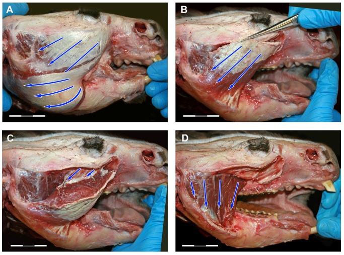
Fig 3. Dissection photos of the masseter muscle in lateral view showing fibre direction.
(A) The most superficial layer of the masseter showing the fibre direction of the internal and external superficial masseter; (B) External superficial masseter removed to reveal the internal superficial masseter; (C) Part of the internal superficial masseter pulled back to reveal the fascia that partially separates the internal superficial masseter at its origin; (D) Internal and external superficial masseter removed to reveal the posterior and anterior deep masseter.

