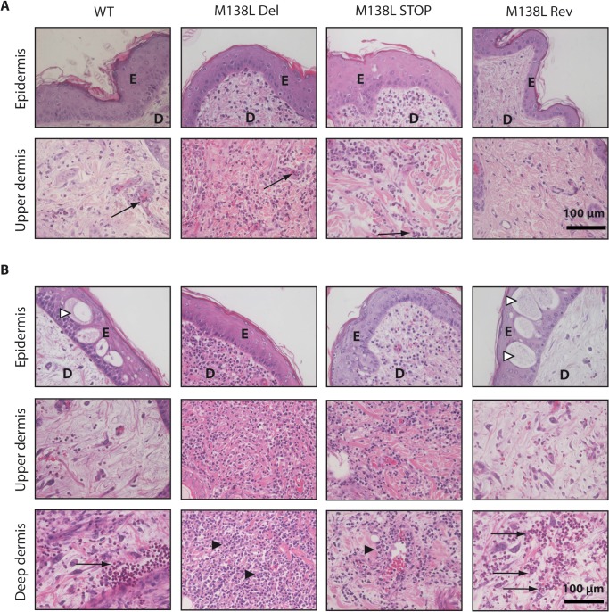
Fig 5. Histological differences at the primary myxoma site from rabbits infected by the Myxoma virus strains.
NZW rabbits were inoculated with 100 PFUs of Myxoma virus WT, M138L Del, M138L STOP or M138L Rev strains in the left flank. A. Haematoxylin-eosin staining at day 4 p.i. B. Haematoxylin-eosin staining at day 9 p.i. These pictures are representative of observations done at least on 4 rabbits. E: epidermis, D: dermis. Black arrows show neutrophils, black arrowheads show mononuclear cells and white arrowheads show ballooning degeneration.

