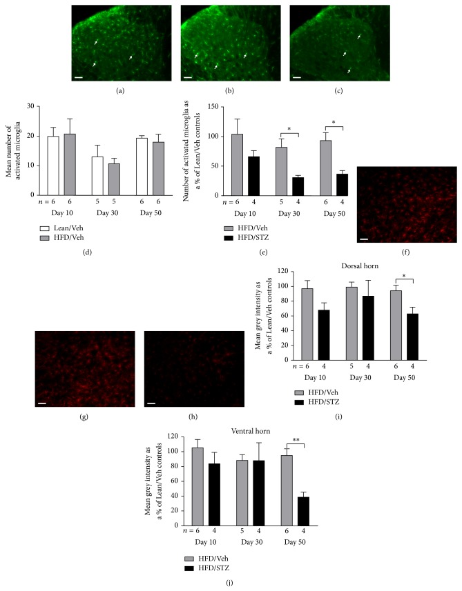
Figure 4.
Effects of the HFD/STZ model on Iba-1 and GFAP staining in the spinal cord. ((a)–(c)) Representative images of Iba-1 staining in the dorsal horn of the spinal cord in a Lean/Veh, HFD/Veh, and HFD/STZ rat, from left to right. Scale bar: 50 μm (20x magnification). Arrows indicate examples of Iba-1 positive cells morphologically identified as activated microglia. (d) Numbers of Iba-1 positive cells morphologically identified as activated microglia in the dorsal horn of the control groups (lean/vehicle and HFD/vehicle). (e) Effects of HFD/STZ on number of Iba-1 positive cells morphologically identified as activated microglia in the dorsal horn expressed as a percentage of the number in the Lean/Veh control group. The numbers of rats per group are shown below the x-axis, and 7 spinal cord sections per rat were processed. Data are mean ± SEM. Analysis was by a Mann-Whitney test: * P < 0.05. ((f)–(h)) Representative images of GFAP staining in the ventral horn of the spinal cord in a Lean/Veh, HFD/Veh, and HFD/STZ rat, from left to right. Scale bar: 50 μm (20x magnification). Effects of HFD/STZ on mean grey intensity of spinal GFAP immunofluorescence in (i) the dorsal horn and (j) the ventral horn, expressed as a percentage of the mean grey intensity in the Lean/Veh control group. The number of rats per group is shown below the x-axis, and 7 spinal cord sections per rat were processed. Data are mean ± SEM, analysis was by a Mann-Whitney test: * P < 0.05, ** P < 0.01.

