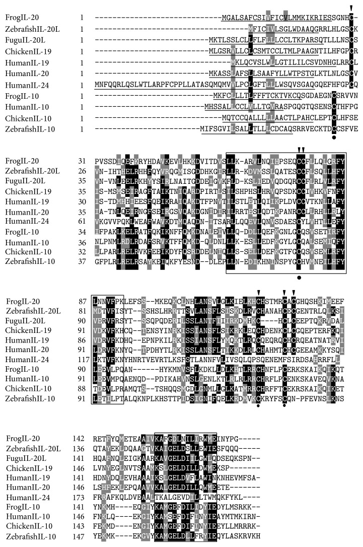
Figure 2.
Multiple alignment of vertebrate IL-10. The multiple alignment was produced using Clustal W, and conserved amino acids were shaded using GeneDoc software. The signal peptides predicted by SignalP 4.1 server were underlined. The conserved IL-10 family signature motifs were boxed. The four conserved cysteine residues existing in IL-10 were indicated by black cycles below the alignment and the six conserved cysteine residues in IL-19/20 were indicated by black arrows above the alignment. The accession numbers for sequence used in this alignment were listed in Figure 6.

