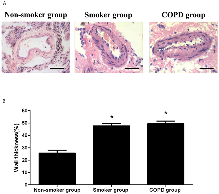
Figure 1. Morphological features of pulmonary artery of nonsmoker group, smoker group, and COPD group.
(A). The lung sections were stained with H&E to evaluate the vessel wall thickness (Scale bars: 20 μm). (B). Pulmonary vessel wall thickness (WT) was expressed as % total diameter in pulmonary arteries. *P < 0.05 versus nonsmoker group.

