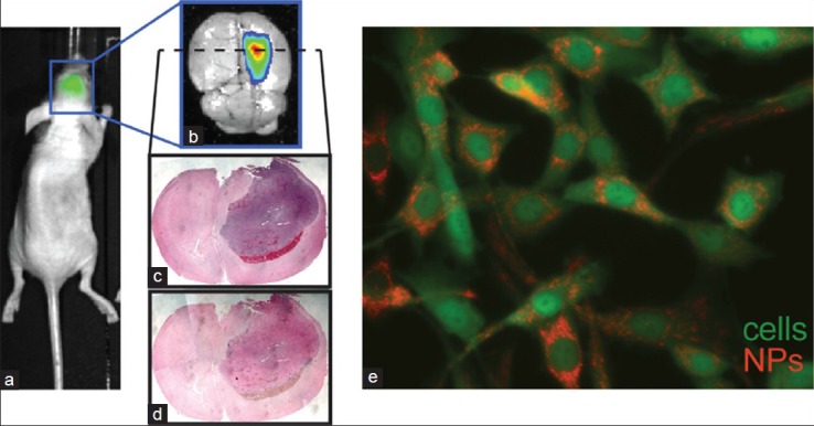
Figure 2.

Targeting of iron oxide nanoparticles to orthotopic C6 glioma xenograft tumors in mice. (a) IVIS bioluminescent imaging of luciferase signal demonstrates tumor location. (b) Fluorescence imaging of red channel (710 nm) shows concentrated presence of cy5.5 fluorophore-labeled NPs within the glioma mass. (c) Hemotoxylin and eosin, and (d) prussian blue/nuclear fast red stained sections of the tumor show accumulation of iron oxide 24 h after injection. (e) Fluorescence microscopy of C6 cells loaded with CTX/cy5.5-bound NPs in vitro
