
Figure 4. Model of protein distribution inside the tegument of HSV-1.
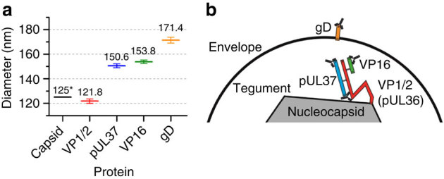
(a) Average diameter of protein layers, error bars are 95% confidence interval obtained from the fit. (b) HSV-1 protein architecture model. This model was built from the diameters obtained using primary labelling and biochemical evidences of interaction sites and antibody-binding sites. *The diameter shown for the capsid was obtained from published EM data5.