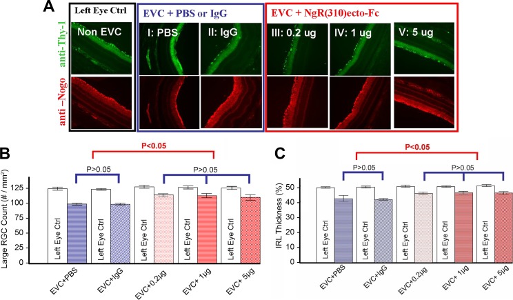
Figure 1.
RGC rescue using rat NgR1(310)-Fc after episcleral vein cauterization. (A) Significant RGC loss was observed in all EVC + treatment eyes (I–V) compared to non-EVC left eyes. The RGC loss in NgR1(310)-Fc treatment groups (III–V) is significantly less than control groups (I and II). (B) Large RGC density (per mm2). Significant reductions (P < 0.05) in large RGCs density were observed in EVC eyes versus non-EVC eyes in all groups. The reduction of large RGC in NgR1(310)-Fc treatment groups (III–V) is significantly (P < 0.05) less than in control groups (I and II). (C) Percentage IRL thickness (% IRL/total retina thickness). Hematoxylin and eosin (H&E) and immunohistology staining demonstrate the significant reduction (P < 0.05) of IRL (RGC and inner plexiform layer [IPL]) thickness in EVC + treatment eyes versus the non-EVC eyes. A significant reduction (P < 0.05) IRL thickness occurs in nontreated EVC eyes (I and II) comparing to NgR1(310)-Fc–treated eyes (III–V). Data are mean ± SEM.

