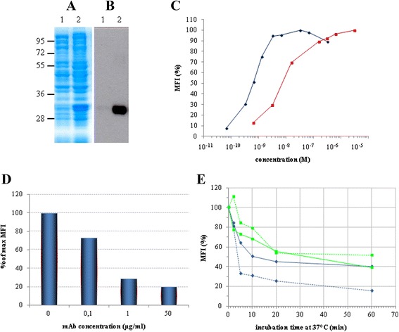
Figure 1.

Expression & characterization of the 4KB scFv. Total lysate of non-induced (lane 1) and IPTG-induced (lane 2) E. coli BL21(DE3) pLys transformed with pET20b(+)4KBscFv were loaded and the expression of the recombinant protein was detected by (A) Coomassie blue staining or (B) Western blot analysis with anti-His antibody. (C) The binding activity of 4KB scFv (red squares) was compared with that of 4KB128 mAb (blue diamonds) by flow-cytometric analysis on Daudi cells incubated at 4°C using increasing amounts of purified 4KB128 mAb or 4KB scFv. (D) The binding of 4KB scFv (50 μg/ml) on Daudi cells is competitively inhibited by increasing concentrations of the parental anti-CD22 mAb pre-incubated with the cells. The scFv-associated fluorescence without competing mAb pre-incubation is taken as the maximal reference MFI. (E) Internalization and stability of the anti-CD22 mAb compared to 4KB scFv. Ramos (light blue) and Daudi (green) cells were stained at 4°C with 30 μg/ml 4KB scFv (continuous line) or 10 μg/ml mAb (dashed line) and subsequently incubated at 37°C for the indicated times, as described in Methods. Red lines indicate the MFI obtained by staining Daudi cells with the scFv (continuous line) and mAb (dashed line) previously incubated at 37°C for the same time lengths as for the internalization experiment. MFI values are plotted as percentage relative to the fluorescence obtained for samples kept on ice.
