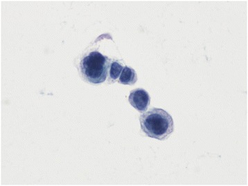
Figure 2.

Presence of tumor cells present in the cerebrospinal fluid. A papanicolaou-stained, liquid-based, thin-layer preparation of cerebrospinal fluid is shown (400×). Tumor cells were of various large sizes, appeared clustered together, and had abnormal ratios of nucleus (dark blue) to cytoplasm (light blue). The features suggested adenocarcinoma cells.
