
Figure 1.
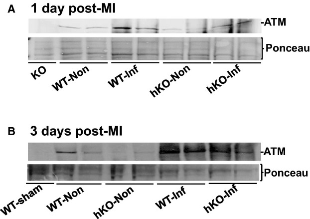
MI increases ATM expression in the heart. LV lysates prepared from WT‐sham, ATM KO, and non‐infarct (Non) and infarct (Inf) LV regions of WT and hKO 1 and 3 days post‐MI were analyzed by Western blot using anti‐ATM antibodies. A, 1 day post‐MI; B, 3 days post‐MI. Protein loading is indicated by Ponceau‐S staining. ATM indicates ataxia telangiectasia mutated; hKO, heterozygous knockout; KO, knockout; LV, left ventricle; MI, myocardial infarction; WT, wild‐type.