
Figure 3.
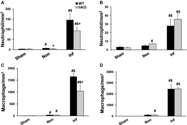
ATM deficiency results in decreased inflammatory cells in the infarct LV region 1 day post‐MI. Cross‐sections of the heart post‐MI were immunostained using anti‐neutrophil (A and B) and anti‐F4/80 (macrophage; C and D) antibodies. The number of immune‐positive cells was quantified using Bioquant Image analysis software. Quantitative analyses of neutrophils 1 (A) and 3 (B) days post‐MI. Quantitative analyses of macrophages 1 (C) and 3 (D) days post‐MI. #P<0.05 vs Sham, $P<0.05 vs Non, *P<0.05 vs WT; n=4 to 6. ATM indicates ataxia telangiectasia mutated; hKO, heterozygous knockout; Inf, infarct; LV, left ventricle; MI, myocardial infarction; WT, wild‐type.