
Figure 4.
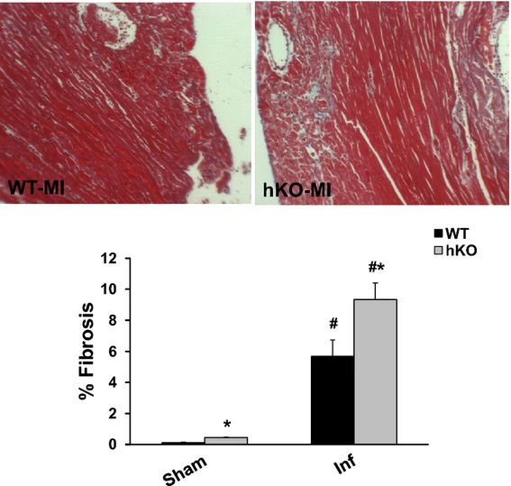
ATM deficiency results in increased fibrosis. Masson's trichrome stained sections of the heart were used for quantitative measurement of fibrosis. Upper panel depicts Masson's trichrome‐stained sections exhibiting fibrosis in WT and hKO hearts 3 days post‐MI. Lower panel exhibits quantitative analysis of fibrosis. #P<0.05 vs Sham, *P<0.05 vs WT‐MI; n=5 to 6. ATM indicates ataxia telangiectasia mutated; hKO, heterozygous knockout; Inf, infarct; MI, myocardial infarction; WT, wild‐type.