
Figure 6.
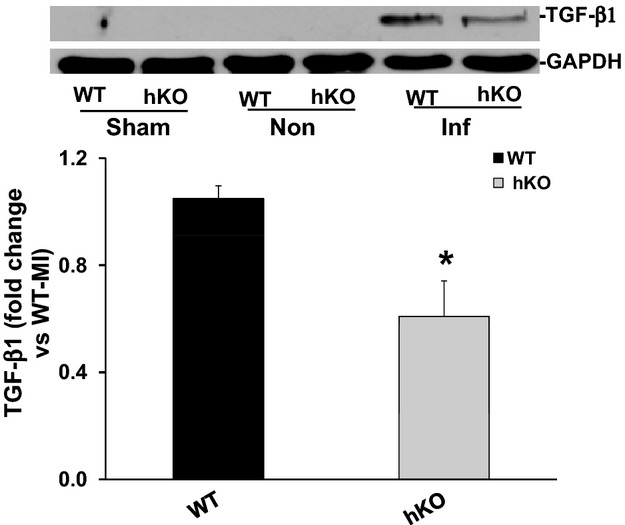
Expression of TGF‐β1. Total LV lysates, prepared from sham and non‐infarct (Non) and infarct (Inf) LV regions 3 days post‐MI, were analyzed by Western blot using anti‐TGF‐β1 antibodies. The upper panel depicts autoradiogram indicating immunostaining for active TGF‐β1 (≈26 kDa band) and GAPDH. The lower panel exhibits quantitative analysis of TGF‐β1 in the Inf LV regions of WT and hKO groups normalized to GAPDH. *P<0.05 vs WT‐Inf; n=8 to 9. hKO indicates heterozygous knockout; Inf, infarct; LV, left ventricle; MI, myocardial infarction; TGF‐β1, transforming growth factor‐beta 1; WT, wild‐type.