
Figure 9.
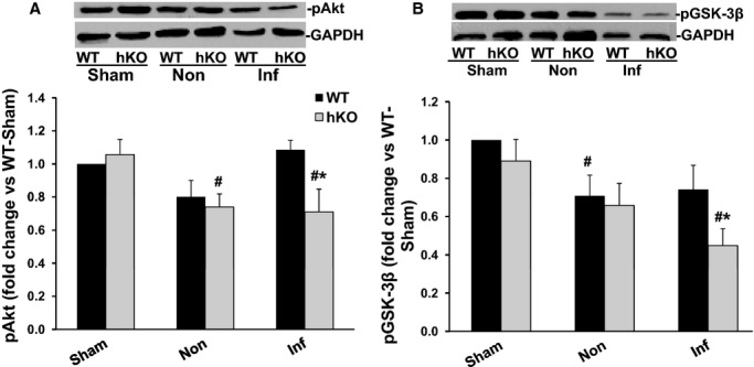
Phosphorylation of Akt and GSK‐3β. Total LV lysates, prepared from sham, non‐infarct (Non) and infarct (Inf) LV regions 1 day post‐MI, were analyzed by Western blot using phospho‐specific antibodies for Akt (ser‐473) and GSK‐3β (ser‐9). The upper panels depict autoradiograms indicating immunostaining for p‐Akt, p‐GSK‐3β and GAPDH. The lower panels exhibit quantitative analyses of p‐Akt (A) and p‐GSK‐3β (B) normalized to GAPDH. #P<0.05 vs Sham; *P<0.05 vs WT‐Inf; n=6. GSK, glycogen synthase kinase; hKO indicates heterozygous knockout; LV, left ventricle; MI, myocardial infarction; WT, wild‐type.