
Figure 10.
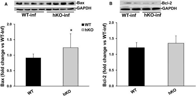
Expression of Bax and Bcl‐2. Total LV lysates, prepared from infarct LV regions 3 days post‐MI, were analyzed by Western blot using anti‐Bax and anti‐Bcl‐2 antibodies. The upper panels depict autoradiograms indicating immunostaining for Bax (A), Bcl‐2 (B) and GAPDH. The lower panels exhibit quantitative analyses of Bax (A) and Bcl‐2 (B) normalized to GAPDH. *P<0.05 vs WT‐MI; n=6 to 8. hKO indicates heterozygous knockout; Inf, infarct; LV, left ventricle; MI, myocardial infarction; WT, wild‐type.