
Figure 2.
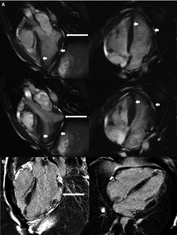
A, Cardiac magnetic resonance imaging 3‐chamber and 4‐chamber views at end‐diastole (top panels) and end‐systole (bottom panels) of a homozygote showing a basal inferolateral aneurysm (long arrow) with additional focal areas of thinning and akinesis in the apex, apical septum, apical anterior, and mid anterolateral walls (short arrows). There were no right ventricular abnormalities. B, Corresponding views showing small regions of late gadolinium enhancement at the edge of the basal inferolateral aneurysm (long arrow).