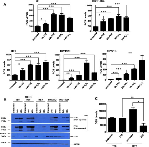
Figure 1. Iron increases ROS in multiple ovarian cell lines, whereas GSH levels diminish upon iron treatment in HEY cells.
(A) T80, T80+H-Ras, HEY, TOV112D and TOV21G cells were assessed for ROS as described in the Experimental section (n=2). (B) T80, T80+H-Ras, HEY, TOV112D and TOV21G cells were treated with 250 μM FAC for 24 and 48 h. Cell lysates were harvested and analysed via Western blotting using the following antibodies: (1) FTH1, (2) CD71, and GAPDH. (n=3). (C) T80 and HEY cells were assessed for GSH levels as described in the Experimental section (n=3).

