
Figure 2. Contribution of IRP1/2 to ferritin and CD71 protein expression upon FAC treatment.
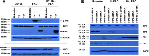
(A) HEY cells were seeded at 250000 cells in each well of a six-well plate. After overnight attachment, the cells were treated for 18 h with U0126 alone, FAC alone or U0126 and FAC combination. Cell lysates were harvested and analysed via Western blotting using the following antibodies: (1) p-ERK, (2) total MAPK, (3) ferritin (FTH1), (4) CD71 and (5) GAPDH (n=2). (B) HEY cells were seeded at 325000 cells in each well of a six-well plate. Following cellular adherence, the cells were treated with two rounds of control non-targeting, IRP1, IRP2 or IRP1 and IRP2 siRNA on sequential days as described in the Experimental section. Iron (250 μM) was then added for 1 or 18 h. Cell lysates were harvested and analysed via Western blotting using the following antibodies: (1) IRP1, (2) IRP2, (3) ferritin (FTH1), (4) CD71 and (5) GAPDH (n=4).