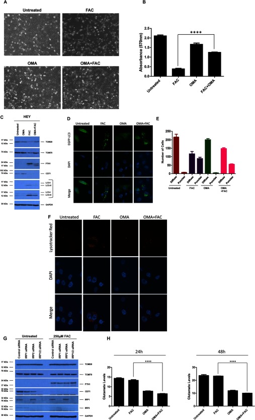
Figure 5. Oxalomalate reverses FAC-mediated loss of OMM proteins and cell death in HEY cells.
(A) HEY cells were seeded at 250000 cells in each well of a six-well plate. Following adherence, cells were treated with FAC (250 μM) alone, OMA (5 mM) alone or FAC (250 μM) in combination with OMA (5 mM) for 48 h. Representative light microscope images were then captured. (B) HEY cells were seeded at 5000 cells per well in a 96-well plate. Following attachment, cells were treated as described in (A), stained with Crystal Violet and dissolved with Sorenson's Buffer followed by quantification at 570 nm. (C) HEY cells were treated as described in (A). Cell lysates were analysed by Western blotting using the following antibodies: (1) TOM20, (2) TOM70, (3) FTH1, (4) CD71, (5) LC3B and (6) GAPDH (n=2). (D) HEY cells were seeded at 250000 cells in each well of a six-well plate. Following adherence, cells were transfected with pEGFP-LC3 and then treated as described in (A) for 24 h. Images were then captured using a confocal microscope and a 60× oil immersion objective lens and representative images are shown (n=2). (E) Quantitative data for results presented in (D) were obtained by counting at least 200 GFP positive cells and assessing the number of cells containing more than 20 punctae (n=2). (F) HEY cells were treated as described in (A) for 24 h. Following treatment, LysoTracker Red was added for 1 h. The cells were stained with DAPI and viewed using a confocal microscope with a 60× oil immersion objective lens. Representative images are shown (n=2). (G) Following cellular adherence, HEY cells were treated with control non-targeting, IRP1, IRP2 or IRP1 and IRP2 siRNA. Iron (250 μM) was then added for 48 h. Cell lysates were harvested and analysed via Western blotting using the following antibodies: (1) TOM20, (2) TOM70, (3) FTH1, (4) CD71, (5) IRP1, (6) IRP2 and (7) GAPDH (n=4). (H) HEY cells were treated as described in (A). Glutamate levels were assessed by harvesting the media supernatant at 24 h (left panel) or 48 h (right panel) following treatment (n=2).

